- Home
- Blog
- Digital dentistry
- How an intraoral scanner works
How does an intraoral scanner work?
Having written about digital dentistry now for several years, I realized I never explored in detail how exactly our intraoral scanner technology works. So I walked into our TRIOS department and asked my colleagues there to explain how a TRIOS intraoral scanner works and in general, how intraoral scanners work.
Intraoral scanner technology explained, step by step
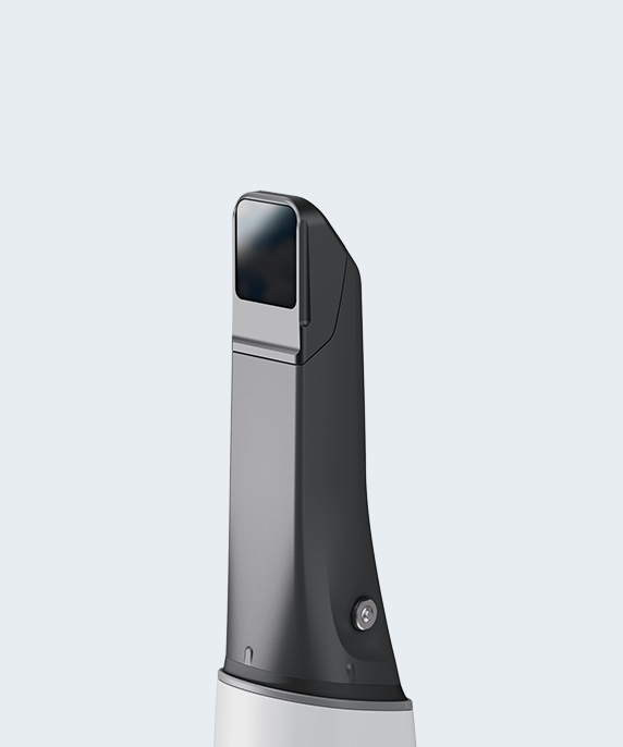
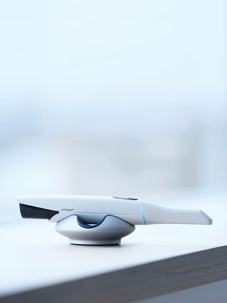
There, I ran into Mads Falkenberg Christiansen, and Gabija Kirsanske. Listening to them describe what goes into creating a 3D scan was akin to sneaking into the wrong lecture by mistake: one on quantum physics. It is simply mind-boggling the amount of technology that gets packed into TRIOS. Especially one as compact as our latest TRIOS 5 wireless intraoral scanner.
In working principle, an intraoral scanner is a camera. Like a camera, it bounces light off an object. The scanner’s lens then redirects the light back to a sensor, a single point to create a sharp image. With these images, it builds a dental model.
An intraoral scanner does this just like a digital camera, but it does it by taking images at incredibly fast speeds while crunching enormous amounts of data. Then, to top it off, the scanner and software stitch together the thousands of images to create a 3D image of an object, which also includes color information.
Digital dental scanning by the numbers
A TRIOS intraoral scanner takes approximately 2,500 images per second. Let that number sink in.
To put the 2,500 images/second in perspective:
- Standard definition video captures 25 frames/images per second.
- High-end cameras take anywhere from 30 to 60 fps/images/sec with some newer models up to 120 fps.
- TRIOS is 2,500 images/frames per second!
From approximately every 100 images taken by TRIOS’ sensor, it creates a sub-scan. A sub-scan is a point cloud or a 3D representation (including colors) of what the scanner sees in front of it.
The size, known as volume, of a sub-scan is comparable to the size of the scanner’s tip window. In TRIOS case, this is around 2x2x2 cm. TRIOS generates some 25 of these sub-scans per second.
Intraoral scanner working principles: data size
The lightning quick image sensor also generates approximately 2.5 gigabytes of data per second. That means the scanner’s hardware and software need to instantly crush the data down to around 40 MBs just to make it manageable to transmit and process it on a PC. If it did not do that, scanning a single arch made up of all these point clouds would probably blow out your practice or lab’s network.
So, while the scanner’s hardware is taking the images, calculating sub-scans, and transmitting them to the PC, the TRIOS software is crunching the data and stitching together the thousands of sub-scans. It is also building up the 3D model of the full dentition, including color and fluorescence textures.
At the same time, it is also finding out where each sub-scan fits on the model and continually updating the model with the new information. In real-time.
These point clouds, organized into triangles, is where the triangle in the 3Shape logo comes from.
You will probably recognize it if you have ever seen one of those black and white images of a scan with all those triangles on it.
While the scanner is busy snapping images, the scanner’s AI is evaluating the images and saying OK, that’s a tongue, we don’t want that, or that’s my finger, we definitely don’t want that. It does this in real-time too.
The scanner is taking all these sub scans and building a digital model of the mouth in 3D and color.
A wireless intraoral scanner’s working principles
For me, this is when things really get interesting. Because, as mentioned, TRIOS is taking these 2,500 images per second but, every single image is 2D, like a camera’s flat image. So how does it turn it into 3D?
Gabija explains, it’s called focus scanning. It’s also what separates TRIOS from a lot of scanners in the market. And why 3Shape has so many patents related to focus scanning.
Without giving away any trade secrets, like a camera, TRIOS has lenses in it. And like a camera, the lens system can focus on different distances. The TRIOS scanner focuses in and out automatically. At the same time, there is a scale that the focus lens is riding on that tracks its position while it moves back and forth on the track. So, for every image taken there is also a corresponding position, and it can calculate a depth associated with that image.
So, when an image is shot, the camera knows that the lens was in this position to get this image in focus and in the next image it was in that position to be able to focus on the image. All of a sudden, we are measuring and comparing depth between the two images.
Teeth are strange objects
But here’s where things get really interesting. As dental professionals know, teeth are strange objects that don’t photograph too well. To be able to take a good image of a tooth, TRIOS needs to use an image and project that image onto the tooth’s surface.
Otherwise, the light would either bounce right off or be soaked into the tooth or pass through it. That is because teeth are weird. They are translucent. Gabija expands on this a little later.
From checkerboard to point clouds
But first, back to TRIOS… the image that it shoots onto the tooth looks like a checkerboard. TRIOS figures out where its lens needs to be to have that projected checkerboard image in focus and from that, it can compute the depth of that image. The checkerboard pattern creates contrast on the tooth, which in turn, helps to compensate for the tooth’s translucency.
Gabija says the checkerboard images enable us to create layers, which are slices of images at different depths. These layers are what the 3D sub-scan is built from.
In her words, “depth information is extracted by measuring the distance to the focal plane with a scale on the lens. The layered images are pooled into creating one of the many point clouds” - those 2.5 GB/sec files that we crush down to 40 MB/sec.
When you think about it, it now makes sense why scanners have difficulty scanning certain areas of the mouth. They not only have to be able to take a picture of a hard-to-reach area, but they also need to measure the depth of that area to create a 3D image.
That means, in the case of TRIOS, bouncing that checkerboard into that hard-to-reach area, getting it in focus and identifying the depth of the shot, and do that, at 2,500 images per second!
Different intraoral scanner types
It’s also what separates the more expensive scanners from the lower cost models, says Mads. The ability of the intraoral scanner manufacturer’s software to process all these images/sub scans varies wildly, especially when it comes to images from interproximal areas.He says that the lower price scanner models struggle with these difficult surfaces. Every scanner in the market can take a digital impression or 3D scan.
But the lower price scanners have a tougher time getting data from hard-to-reach areas in between teeth, distal surfaces, and undercuts, for example.
The interesting optical properties of teeth
So back to teeth - your favorite subject. In Gabija’s words, “teeth are exotic surfaces to scan.”
She continues, “teeth have very interesting optical properties. When the tooth is illuminated with uniform lighting, it exhibits a consistent reflection due to its smooth surface and uniform color. Furthermore, when light is projected onto a tooth some of the light gets scattered inside the tooth because the tooth is semi-translucent. To be exact, tooth enamel is semi-translucent.
For TRIOS, to build a 3D model, we need to scan the surface to match the tooth not the inside of the tooth.”
By projecting the checkerboard image onto the tooth, TRIOS tackles these two challenges. First, it uses the pattern (not necessarily with polarized light) to create that contrast on the tooth’s surface.
Second, by using light polarization, it enhances the signal from the surface of the teeth.
Intraoral scanner accuracy
This also makes perfect sense. Because we are creating a 3D image, and the depth needs to be precise and measured from the tooth’s surface. If you are creating a crown based on a scan, then it better obtain the measurement data from the outside of the tooth, as opposed to a millimeter from within the tooth because the tooth happened to be translucent. Now we can see why translucency can impact intraoral scanner accuracy.
Intraoral scanner types: powder versus non powder
In the old days when scanners used powder it was a lot easier. The powder eliminated the translucency. The problem is powder is a pain and nobody likes it. If you are interested in learning more about the optical characteristics of natural of teeth, the article by Dr. James Fondriest gives more information.
Because teeth are translucent, and not transparent, 3Shape employs a different technology to scan a tooth’s surface. It’s also why the art of “characterizing” dentures or any restoration for that matter, is such a unique skill.
Intraoral scanner technology: polarization
The technology TRIOS uses to combat translucency is called polarization, and it is where Gabija’s expertise comes in. She explains that polarization technology, which is also used in many digital cameras, is what helped TRIOS make powdered scanning obsolete way back when.
Without getting into too much detail, light reflects off or within a tooth in three ways: specular, surface scattering, and subsurface (volume) scattering.
The reflection of the light from TRIOS on the tooth becomes random or unpolarized by these three types of waves. Again, we could cure this by using powder and making the tooth completely opaque, but polarization technology and engineers like Gabija devised an alternative. The technology basically allows TRIOS to differentiate the polarization fingerprint of the light signal arriving predominantly from the surface of the tooth.
Unfortunately, Gabija has requested that we do not talk anymore about TRIOS’ application of polarization technology. In her words, “we do have to have secrets.”
Final thoughts on choosing between intraoral scanner manufacturers
- Check-out clinical studies
You will want to focus on intraoral scanner accuracy. “There are plenty of independent studies available. The 3Shape website has a library of clinical studies for your review.” - Test models
Test various scanner models at shows and see which brand feels most comfortable in your hand and has the easiest software to work with. Dr. Mark McOmie created a terrific model for evaluating intraoral scanner manufacturers. - Test in vivo
Most importantly, test the scanner on a mouth in vivo. “Scanning plastic or acrylic models is fine for checking the feel, but you really need to see how the scanner reacts to saliva, hard to reach areas, speed, and comfort.
Remember, every scanner in the market can scan a model. Not every scanner can completely scan a mouth.”
Explore the future of dental impressions with us