
Most practitioners recommend that you sit directly behind your patient when intraoral scanning. Australia’s Leif Svensson, who might just be closing in on the Most Intraoral Scans Taken career record, says there’s another way. You should stand and face your patient. Read why.
I have been around a little while now and I can safely say that I have seen all almost every conventional impression technique, type of material, as well as impression trays known to mankind.
I have seen impressions pour into laboratories by the truckload over the years. Some very good and some not so good.
How an analog impression study helped me
Two years before my digital journey began, I ran a research project across a large catchment of dental practitioners. The study included general dentists, specialists (Special Pros, Ortho, Perio) and Denturists. My goal was to work out which practitioner category was the best impression takers and why.
A set of evaluation markers and parameters were determined. It’s probably no surprise that the technical staff evaluating some 400 anonymous impressions determined that the best impression takers for extensions and landmarks in this study were, Prosthodontists, Denturists and Orthodontists (I won’t say in which order!).
From the collected data, I determined what these three categories of practitioners had in common, and why they consistently delivered such great conventional impressions.
Here is what I learned on taking the best dental impressions
1. Practice. The practitioner had taken lots and lots of impressions. At least three per day and on most days in an average week.
2. Patient anatomy was quickly analyzed before impression was taken (tray size, opening size, tongue compliance, other anatomical factors in motion).
3. The mix of impression material generally had a thicker viscosity to gain full extension records. These great extensions were achieved through muscle and soft tissue movements.
4. Patient Posture. The patient was in most cases sitting in an upright position for the impression.
5. Both hands were engaged in impression taking. One to insert the tray and one to manipulate the anatomy and muscle trim etc.
6. The practitioner was standing and generally, in frontward facing position relative to the patient for the impressions. Especially lower impressions.
Applying conventional impression-taking best practices to intraoral scanning
FAST Forward a few years and there I was, learning how to intraoral scan very difficult partial and fully edentulous arches. I thought to myself, what could I learn from these amazing impression takers and which of these techniques could I repurpose, digitalize, and then use in my digital record taking bag of tricks?
Ironically, every single one of the above markers translated directly to digital impression taking. Well, almost every one: impression viscosity was replaced by getting yourself the best possible scanner! I’d like to tell you how I managed to digitalize the tips and tricks on conventional impression-taking from the very best in the business, and how these principles help me scan like I do now.
"The key to becoming confident with an intraoral scanner is practice,” Leif Svensson
6 steps to becoming an intraoral scanning pro
The key to becoming confident with an intraoral scanner is practice. Muscle memory is key. Doing as many scans as you can is critical to scan ‘body building’ it, as I elaborated on in my previous blog post on my top 10 learnings for intraoral scanning.
1. Start with a backup.
When you are just starting to scan, I recommend that you first, take a traditional impression. Take the pressure off yourself. Then DO NOT tell the patient what you are doing with the scanner. Just say “I’m just going to take a few photos”. Do this as much as you can until you are confident to scan without the alginate back up! Then say goodbye to the yucky stuff.
2. Analyze your patient.
This may seem a bit weird, but I’m analyzing my patient from the time I walk them from the waiting room and seat them into the chair. Look at the size of the opening, check what happens when you say move your tongue right (does it go left!?). Are there any other anatomical factors that change in motion? How does the intra coronal arch react to opening and closing? Does saliva start to pool as your scanner gets close to the mouth? With practice you will do this very fast. Second nature even. Create a set of quick- fire evaluation parameters that will help you formulate your approach to the IO scan.
3. The technology mix.
Ok this is one thing you don’t have to guess or practice. In the digital world, the mix is just getting yourself the best scanner you can afford with the right studio apps for your practice treatment bag of tricks.
4. Sit upright.
I have absolutely adopted the approach that a patient sitting upright is the best way to capture a great scan. It is doubly important for bite taking for many reasons.
5. Support your scanner.
Supporting your dental scanner with your non grip hand is critical. Use your index finger of the opposite hand to touch a tooth or sulcus as an anchor then the thumb of that free hand becomes the best scanner head support.
6. Frontward face!
Lastly, the case to frontward face! I was a little scarred when training dentists over the years to tell them to get out from the back seat and into the driver’s seat of the scanner process (so to speak).
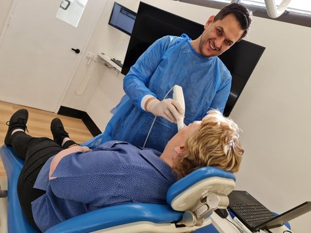
The case to forward face when intraoral scanning
To stand in front of your patient provides some very big advantages. Firstly, you are firmly grounded with full visibility of your screen as well as your patient. Front facing means full access to hand positions only possible in this posture. By standing in front of the patient you can also react to the oral environment quickly. You can take visual cues from your patient if something is uncomfortable, for example. The advantages in complex scanning are immense.
Why not see what analog protocols you can digitalize? There is very good chance it will be world first as we work together to provide the optimum experience for our patients.
Why is it still so uncommon to sit in front?
The reason is because dentists are so used to sitting behind a patient. So, it's easier to just keep that same posture as they are generally still sitting, and patient still reclined. When conducting training, I used to be scared to tell dentists to stand up. But I now realize that more than half of the issues they have are generally solved with this very simple tip (and some practice). We did a lot of filming to document this, and the films have confirmed the benefits of frontward scanning.
And so, I rest my case to Frontward Face!
We’ve asked experience practitioner Dr. Christopher Ho to walk us through how he uses his intraoral scanner for a range of activities, from monitoring tooth wear, performing smile design, diagnosing caries, planning implants and analyzing occlusion.