- Home
- Blog
- Digital dentistry
- Intraoral scanners
Intraoral scanners in dentistry: definition, how they work, and clinical impact
An intraoral scanner is a digital dental device used to capture 3D images of teeth and oral structures. It replaces traditional impressions with digital scans that can be used for diagnosis, treatment planning, and the production of dental restorations.
What is an intraoral scanner?
Intraoral scanners are a type of digital scanners in dentistry used to capture 3D images of teeth and surrounding oral structures. They provide a digital alternative to traditional impression methods by creating accurate intraoral scans that can be used for diagnosis, treatment planning, and restoration design. In practice, this includes designing and producing restorations such as crowns, bridges, veneers, and orthodontic aligners based on digital models of the patient’s teeth.
While different types of intraoral scanners vary in features and software, they all work by capturing detailed 3D data directly from the patient’s mouth. This can reduce the need for physical impressions and enables faster, more consistent handling of clinical data.
How does an intraoral scanner differ from traditional impression methods?
Intraoral scanners capture digital 3D data directly from the patient’s mouth, while traditional impressions use physical materials to create molds. Digital scans can be processed, stored, and shared more efficiently, reducing the number of manual steps.What is an intraoral scan used for?
An intraoral scan is used for diagnosis, treatment planning, and the design and production of dental restorations such as crowns, bridges, and aligners. It also supports communication between clinics and laboratories through digital data exchange.
Where intraoral scanners fit in the digital dentistry ecosystem
Intraoral scanners are a core component of digital dentistry workflows, used to capture 3D data of teeth and oral structures. This data forms the starting point for a digital process that connects clinical practice with laboratory production.
Once captured, intraoral scans are used in CAD/CAM systems for treatment planning and restoration design 1. The data can then be transferred digitally to labs or in-house production systems for manufacturing using milling or 3D printing.
By replacing physical impressions with digital data, intraoral scanners can support a more connected workflow between clinics and labs, supporting more efficient and consistent treatment processes.
How do intraoral scanners work?
An intraoral scanner works by capturing a series of images of the teeth and oral structures using a handheld scanning device. A light source is projected onto the area being scanned, and imaging sensors record the reflected data.
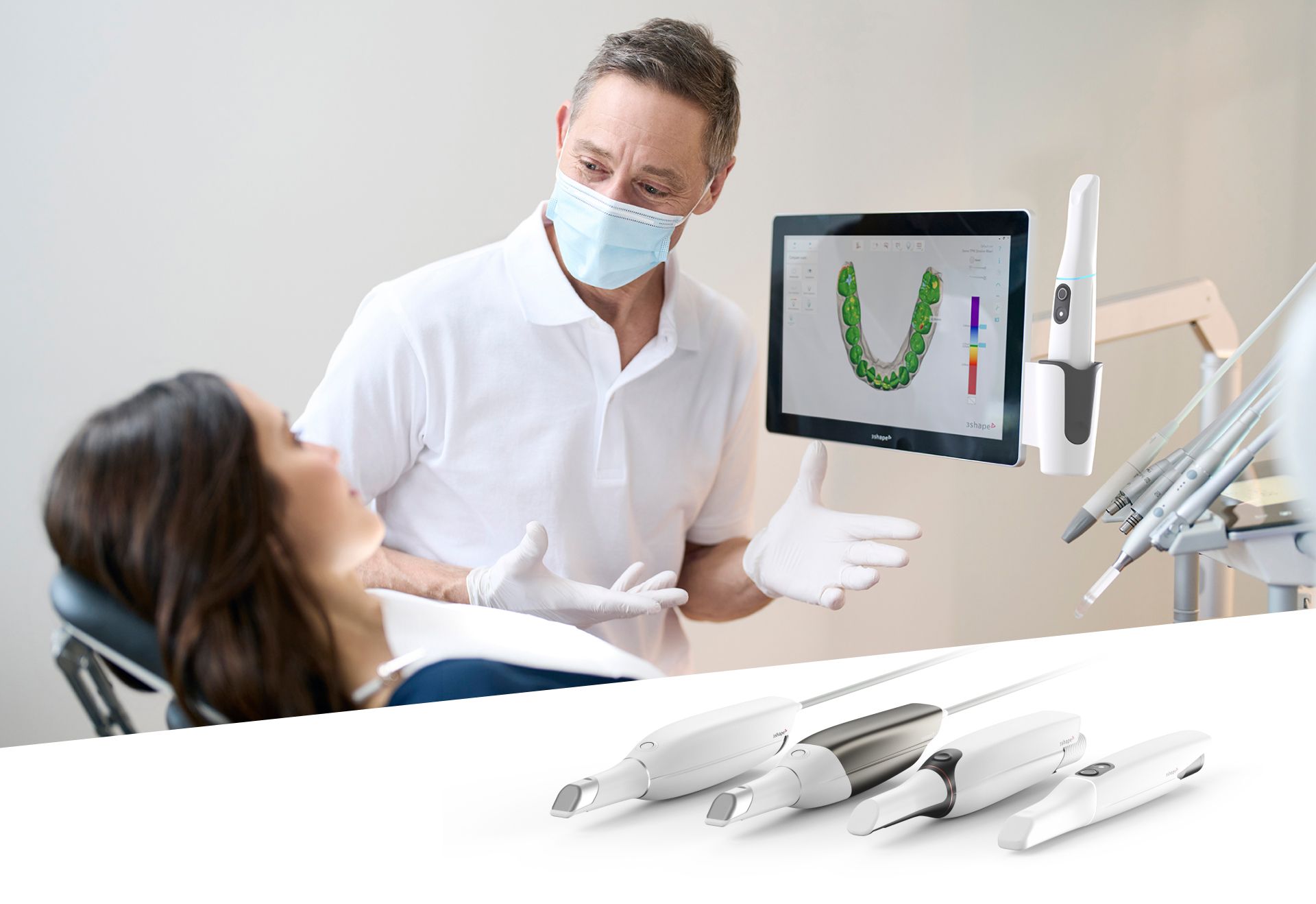
These images are processed by scanning software to create a detailed 3D digital model of the patient’s teeth and gingiva. The captured data is converted into a digital surface model, typically in STL format, which can be used in CAD/CAM workflows. The model is generated in real time and displayed on screen as the scan is performed, as shown in this example of high speed intraoral scanning.
This digital model can then be used in CAD/CAM systems for treatment planning, restoration design, and manufacturing, forming the basis of a digital workflow.
As a result, some manual steps may be reduced, turnaround times may be shorter, and coordination between clinics and labs may be improved.
For a more detailed explanation of the underlying optical and digital technologies, see our guide on how intraoral scanners work.
How do intraoral scanners capture 3D data?
Intraoral scanners use optical technology to capture multiple images of teeth and soft tissues. These images are processed and aligned by software to generate a 3D digital model in real time.
Intraoral scanners vs traditional impressions
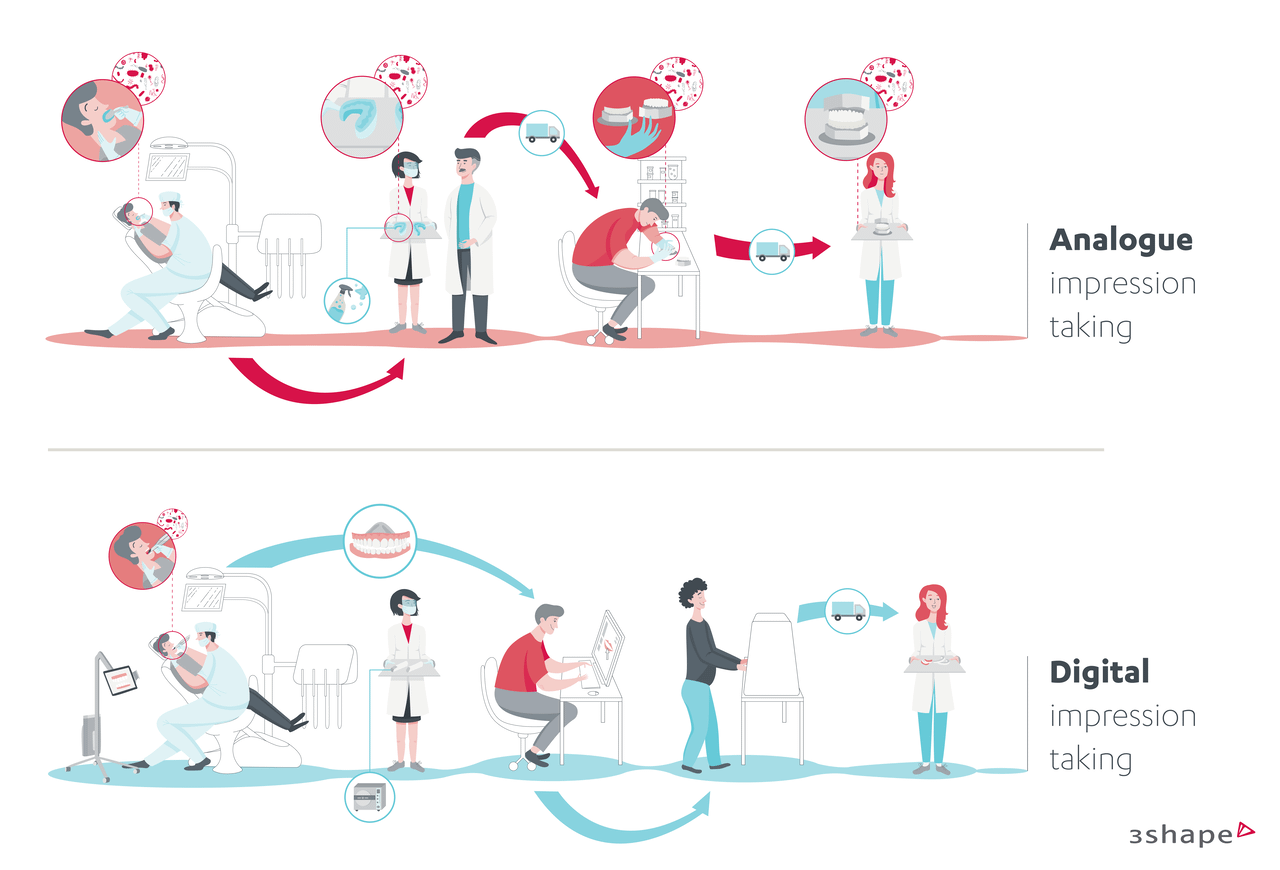
Intraoral scanners and traditional impressions differ in how dental data is captured, processed, and used in clinical workflows. Traditional methods rely on physical materials to create molds, while intraoral scanners capture digital 3D data directly from the patient’s mouth.
With traditional impressions, multiple manual steps are required, including tray selection, material handling, and model creation. Each step introduces the potential for distortion or error. In contrast, intraoral scanning reduces the number of steps by capturing and transferring data digitally.
Digital impressions can be processed, stored, and shared more easily between clinics and labs. This may support more efficient turnaround times and more consistent handling of clinical data compared to conventional impression workflows. For further evidence, you can explore clinical studies comparing digital and conventional workflows.
The key differences between intraoral scanners and traditional impressions are summarized below:
| Feature | Intraoral scanner | Traditional impression |
|---|---|---|
| Material | Digital data | Physical impression material |
| Accuracy | High precision 3D data | High precision. Risk of distortion during impression and casting. |
| Patient comfort | Generally higher | Often less comfortable |
| Data storage | Digital files | Physical models |
What is the difference between intraoral scanners and traditional impressions?
Intraoral scanners capture digital 3D data directly from the patient’s mouth, while traditional impressions rely on physical materials to create molds. Digital workflows reduce manual steps and allow data to be processed, stored, and shared more efficiently.
Clinical applications of intraoral scanners
Intraoral scanners are used across a range of dental treatments where digital impressions are required. The captured 3D data supports diagnosis, treatment planning, and the design of dental restorations and appliances.
Common clinical applications include:
- Crown and bridge restorations
- Implant planning and restoration workflows
- Orthodontic treatment planning and aligner production
- Denture workflows, including digital scan for dentures
- General restorative and prosthetic treatments
In each case, intraoral scanning provides the digital input used in CAD/CAM design and manufacturing within a digital dentistry workflow.
Benefits of intraoral scanners in dental practice
Intraoral scanners affect how treatments are planned, delivered, and experienced in daily practice. By replacing physical impressions with digital intraoral scans, they can support more consistent clinical workflows and improve how data is handled across the treatment process.
Clinical accuracy and treatment outcomes
Digital intraoral scanning supports accurate data capture, which can improve treatment planning and outcomes 2. The clinical impact of intraoral scanning depends on how accuracy is defined and measured in practice, particularly in relation to treatment outcomes.
This is reflected in the following clinical outcomes:
- High-precision 3D data can reduce the risk of distortion compared to physical impressions, with studies showing that digital impressions can be more accurate than conventional methods 2
- More consistent input for CAD/CAM design and manufacturing
- May reduce the need for remakes due to improved fit and accuracy 2
For a deeper look at intraoral scanner accuracy and its impact on treatment quality, download the ebook “Because quality treatment matters to me.”
You can also explore our library of clinical studies on intraoral scanning accuracy and outcomes.
Workflow efficiency and time savings
Intraoral scanners can streamline clinical and laboratory workflows by reducing some manual steps and enabling faster transfer of clinical data.
This is reflected in the following workflow improvements:
- May reduce steps such as impression handling, shipping, and model preparation
- May support faster turnaround times between clinics and labs
- Can improve coordination through digital data exchange
Patient experience and communication
Digital intraoral scanning can improve the patient experience with procedures often reported as more comfortable and easier to understand as in this clinical review of intraoral scanner use and patient preference. 3
This is reflected in the following patient-related outcomes:
- More comfortable than traditional impression materials 3
- Reduced gag reflex and discomfort during scanning 3
- Real-time visualizations that help explain treatment plans
How to choose an intraoral scanner for your practice
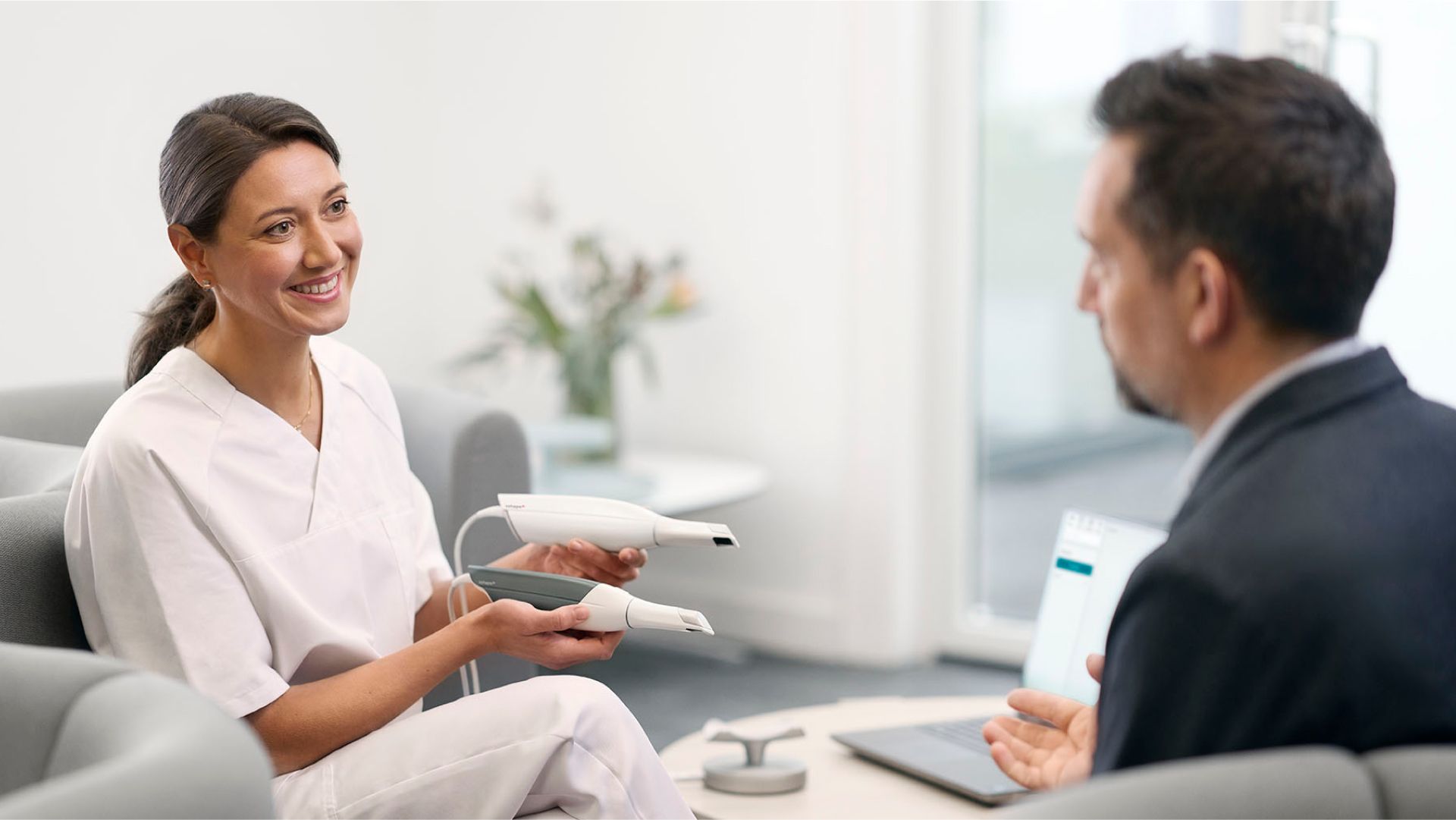
Choosing an intraoral scanner depends on your clinical needs, workflow requirements, and how you plan to use digital impressions in your practice. Different dental digital scanners offer varying features, performance levels, and integration capabilities.
Some brands are relatively new to intraoral scanning technology, while others have developed multiple generations of devices over time. These more established systems often reflect ongoing improvements in performance, usability, and design, including recognition through award-winning products.
For an example of intraoral scanner systems used in digital dentistry, you can explore TRIOS intraoral scanners.
When comparing intraoral scanners, consider the following factors:
- Scanning speed and ease of use during clinical procedures
- Accuracy and consistency of intraoral scans
- Device design, size, and ergonomics
- Software capabilities and integration with CAD/CAM systems
- Total investment, including initial cost and ongoing expenses
Evaluating these factors can help you select a digital intraoral scanner that fits your workflow and supports your treatment needs.
Cost considerations: intraoral scanner price and ROI
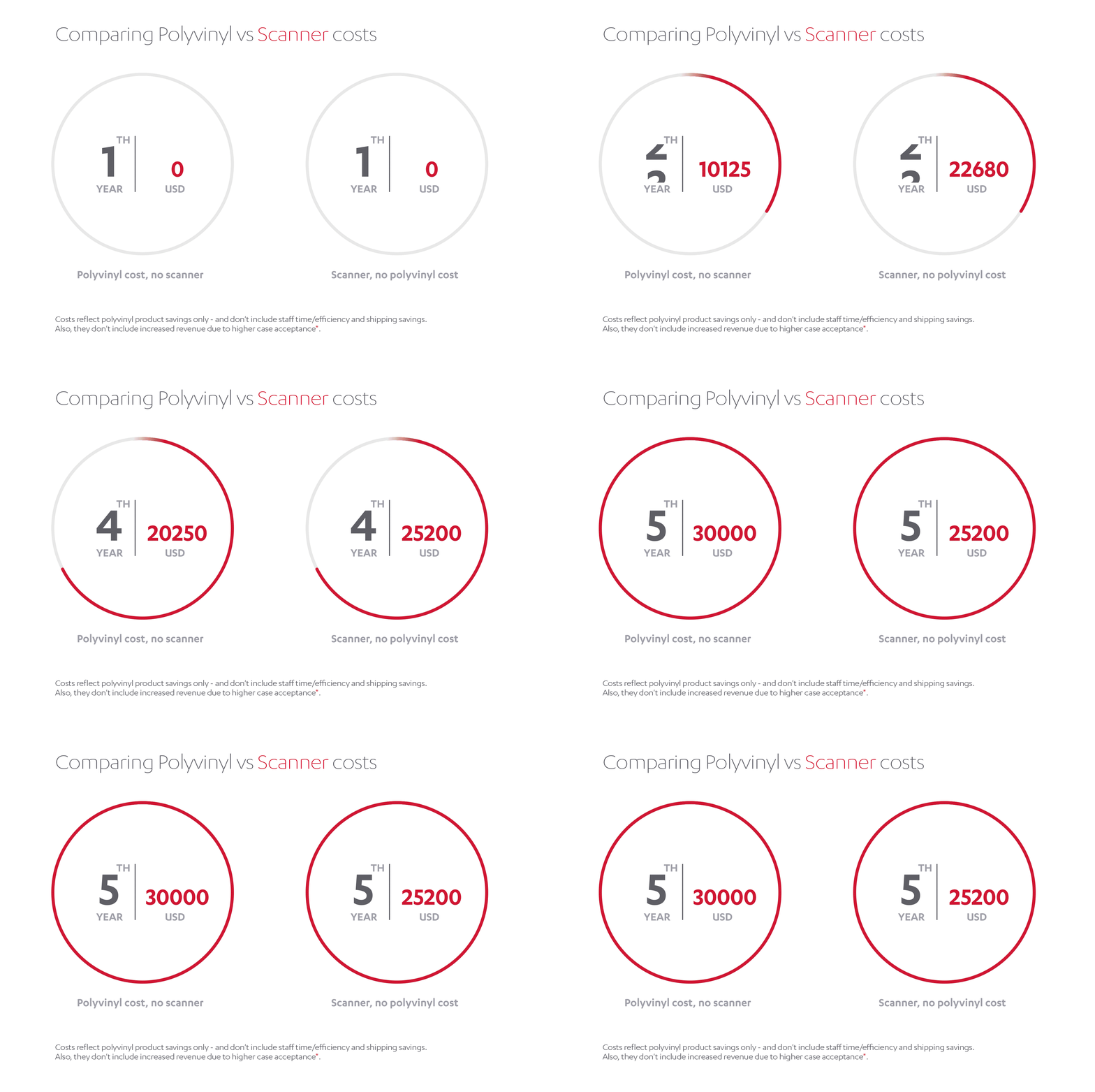
The cost of an intraoral scanner depends on the system, features, and software included. When evaluating price, it is important to consider both the initial investment and the potential return on investment (ROI) over time.
This can be assessed through the following factors:
- Reduction in costs related to impression materials, shipping, and model handling
- Time savings in clinical and administrative workflows
- Improved utilization of staff and resources
- Increased efficiency in case handling and turnaround times
By replacing manual steps with digital processes, intraoral scanners may improve operational efficiency and contribute to offsetting initial costs over time as demonstrated in this comparison of analog and digital impression workflows. For a more detailed estimate, you can use the 3Shape ROI Calculator to evaluate potential savings for your practice.
Future trends in intraoral scanning technology
The use of intraoral scanners has increased as digital dentistry has become more widely adopted. What was initially limited to early adopters is now becoming more common in dental practices.
This shift is reflected in the following developments:
- Increased adoption of digital intraoral scanners across clinical workflows
- Broader use in restorative, implant, and orthodontic treatments
- Greater reliance on digital data for treatment planning and production
- Continued integration with CAD/CAM and digital dentistry systems
Summary
Key takeaways:
- Intraoral scanners are digital dental devices used to capture 3D data of teeth and oral structures.
- They replace traditional impressions with digital intraoral scans that support diagnosis, digital dental treatment planning workflows, and restoration processes.
- Digital intraoral scanning can support more consistent data handling and workflow efficiency within CAD/CAM systems 1
- They are used across a range of treatments, including crowns, bridges, implants, orthodontic aligners, and dentures
- Intraoral scanning can improve patient comfort and communication during treatment 3 and support accurate clinical outcomes 2
For more information, explore these intraoral scanner articles.
Resources
- FDI World Dental Federation: CAD/CAM Dentistry. ADOPTED by FDI General Assembly August, 2017 in Madrid, Spain. https://www.fdiworlddental.org/cadcam-dentistry
- Chandran, Sachin & Jaini, JL & Babu, Anna & Mathew, Anil & Keepanasseril, Arun. (2019). Digital Versus Conventional Impressions in Dentistry: A Systematic Review. JOURNAL OF CLINICAL AND DIAGNOSTIC RESEARCH. 13. 10.7860/JCDR/2019/38494.12756.
- Christopoulou I, Kaklamanos EG, Makrygiannakis MA, Bitsanis I, Perlea P, Tsolakis AI. Intraoral Scanners in Orthodontics: A Critical Review. Int J Environ Res Public Health. 2022 Jan 27;19(3):1407. doi: 10.3390/ijerph19031407. PMID: 35162430; PMCID: PMC8834929.