- Home
- Blog
- Digital dentistry
- Digital Impressions
Digital impressions in dentistry: definition, process, accuracy, and clinical use
A digital impression is a 3D virtual model of a patient’s teeth and oral tissues created using an intraoral scanner or by digitizing a physical model. The system captures surface data from the oral structures and processes it into a digital file that represents the patient’s dentition. Digital impressions are part of the broader digital dentistry workflow and are used for diagnosis, treatment planning, and the design of dental restorations.
Digital impressions are increasingly used as an alternative to traditional impression methods in modern dental practice. Instead of using physical materials, clinicians capture digital data directly from the patient or by scanning a physical model.
This shift supports faster workflows, more efficient data handling, and improved integration with digital systems such as CAD/CAM. Understanding how digital impressions are created and used helps clarify their role in digital dentistry and treatment planning.
The time required to capture a digital impression can decrease significantly with operator experience, as shown in an in vivo study of intraoral scanning performance 1.
What is a digital impression in dentistry?
A digital impression in dentistry is a digital representation of a patient’s teeth and oral tissues that replaces the need for physical impressions. It is created by capturing surface data either directly from the patient using an intraoral scanner or indirectly by scanning a physical model.
Digital impressions are widely used in restorative workflows, including the design and fabrication of dental prostheses 2.
Unlike traditional impressions, which use soft materials to capture the shape of the teeth, digital impressions capture and store this information as a 3D file. This file represents the geometry of the teeth and gingiva and can be viewed, analyzed, and reused without degradation.
Digital impressions have the following characteristics:
- Captured directly from the oral cavity or from a digitized physical model
- Stored as 3D digital files (commonly STL or PLY formats)
- Used for diagnosis, treatment planning, and restoration design
- Easily shared between clinics and laboratories without physical transport
- Integrated into CAD/CAM and digital dentistry workflows
How do digital impressions work?
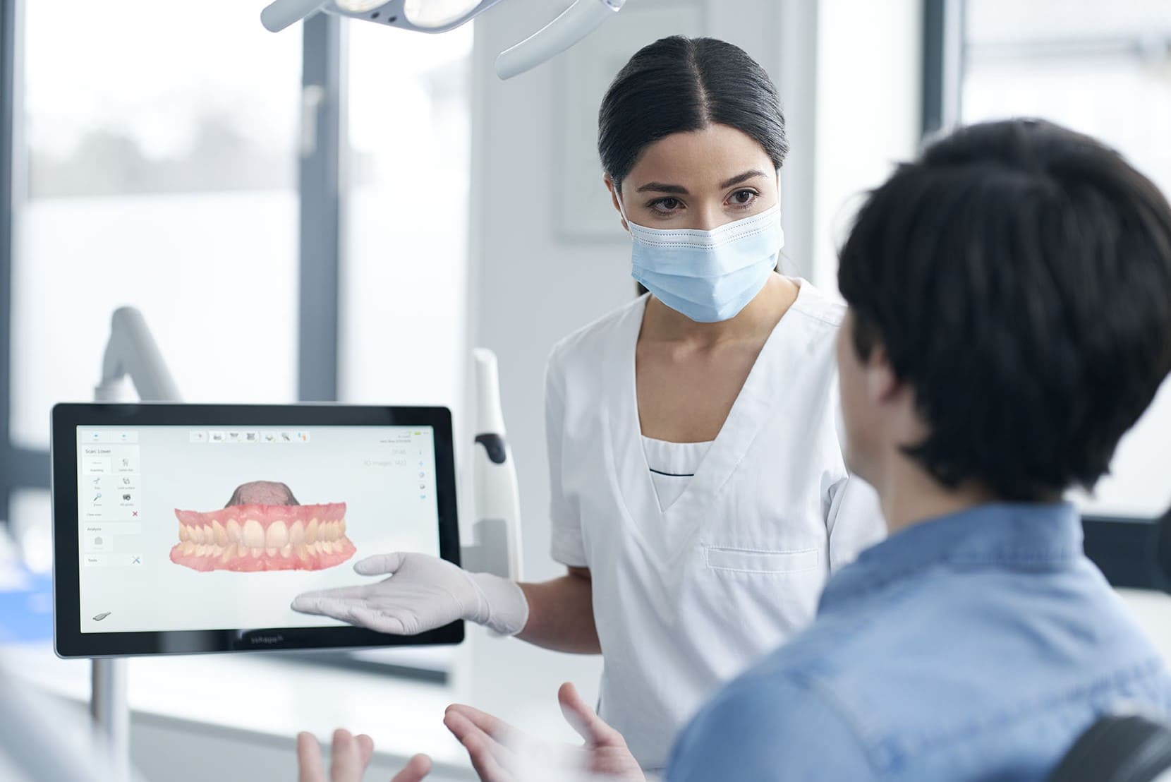
Digital impressions refer to the clinical result of intraoral scanning, not the hardware itself. They represent the outcome of the scanning process rather than the scanning device itself. The process involves capturing surface data from the teeth, processing that data in real time, and generating a digital model that can be used for diagnosis and treatment.
The workflow follows three main stages: optical capture, data processing, and model creation.
Step 1: Optical capture of oral structures
The process begins with capturing surface data from the teeth and surrounding tissues using an intraoral scanner. The device is moved across the dental arches in a continuous motion, capturing images from multiple angles and positions.
Light is projected onto the teeth, and sensors capture how it reflects from the surfaces. This allows the system to map the geometry of the teeth and gingiva.
The result of this step is a series of images representing the scanned areas of the oral cavity.
Step 2: Real-time data processing
The captured images are processed by software in real time. The system aligns and stitches the images together to create a continuous digital representation of the scanned area.
During this stage:
- Overlapping data is aligned and merged
- Surface geometry is reconstructed
- Irrelevant or inconsistent data is filtered out
This results in a structured 3D dataset that represents the shape and position of the teeth.
Step 3: Creation of a digital dental impression
Once the data has been captured and processed, the system generates a complete 3D digital model of the patient’s dentition.
This model is converted into a standard file format, such as STL or PLY, which can be used in CAD/CAM systems for treatment planning, appliance design, and manufacturing.
The digital impression can be stored, reused, and shared across clinical and laboratory workflows.
Process summary
Digital impressions are created by capturing surface data from the teeth, processing it into a structured 3D model, and exporting the file for use in diagnosis, treatment planning, and dental restoration workflows.
How accurate are digital impressions?
Digital impression accuracy refers to how closely a digital scan represents the true shape and position of teeth and oral structures. It is typically evaluated using two parameters: trueness (how close the scan is to the actual geometry) and precision (how consistent repeated scans are).
Clinical evidence shows that digital impressions can achieve accuracy comparable to, and in some cases exceeding, traditional impressions, depending on the clinical indication and scanning conditions, with studies reporting comparable or, in some cases, higher accuracy than traditional impressions 3.
Accuracy is influenced by several factors, including scanning technique, operator experience, accessibility of intraoral areas, presence of saliva or soft tissue movement, and how the software processes and stitches the captured data.
Overall, digital impressions provide consistent and reliable results in many clinical scenarios.
Limitations of digital impressions
Digital impressions are widely used in clinical practice, but they are not without limitations. Certain clinical conditions and practical factors can affect how easily and accurately a digital impression can be captured.
One limitation is the capture of subgingival margins. When preparation margins extend below the gum line, it can be difficult for the scanner to access and record these areas clearly without proper tissue management.
Edentulous and full-arch cases can also present challenges. The lack of stable reference points, combined with movable soft tissue, can make it harder to maintain accuracy across larger scan areas. In response, clinicians have developed specific scanning protocols and techniques for these cases, including corrective impression approaches and strategies for scanning fully edentulous patients 4.
Digital impression workflows may also involve a learning curve. Scanning technique, path, and operator experience can influence the quality of the result, especially in more complex cases, as shown in a study 1 on intraoral scanning performance.
In addition, the initial investment in equipment and software can be a barrier for some practices. Implementing digital impression systems requires not only hardware, but also training and integration into existing clinical workflows.
Despite these limitations, ongoing improvements in scanning technology and software continue to expand the range of clinical situations where digital impressions can be used effectively.
Digital vs traditional impressions
Digital and traditional impressions differ in how dental data is captured, processed, and used within clinical workflows. While both methods aim to reproduce the geometry of the teeth and oral tissues, they rely on fundamentally different approaches.
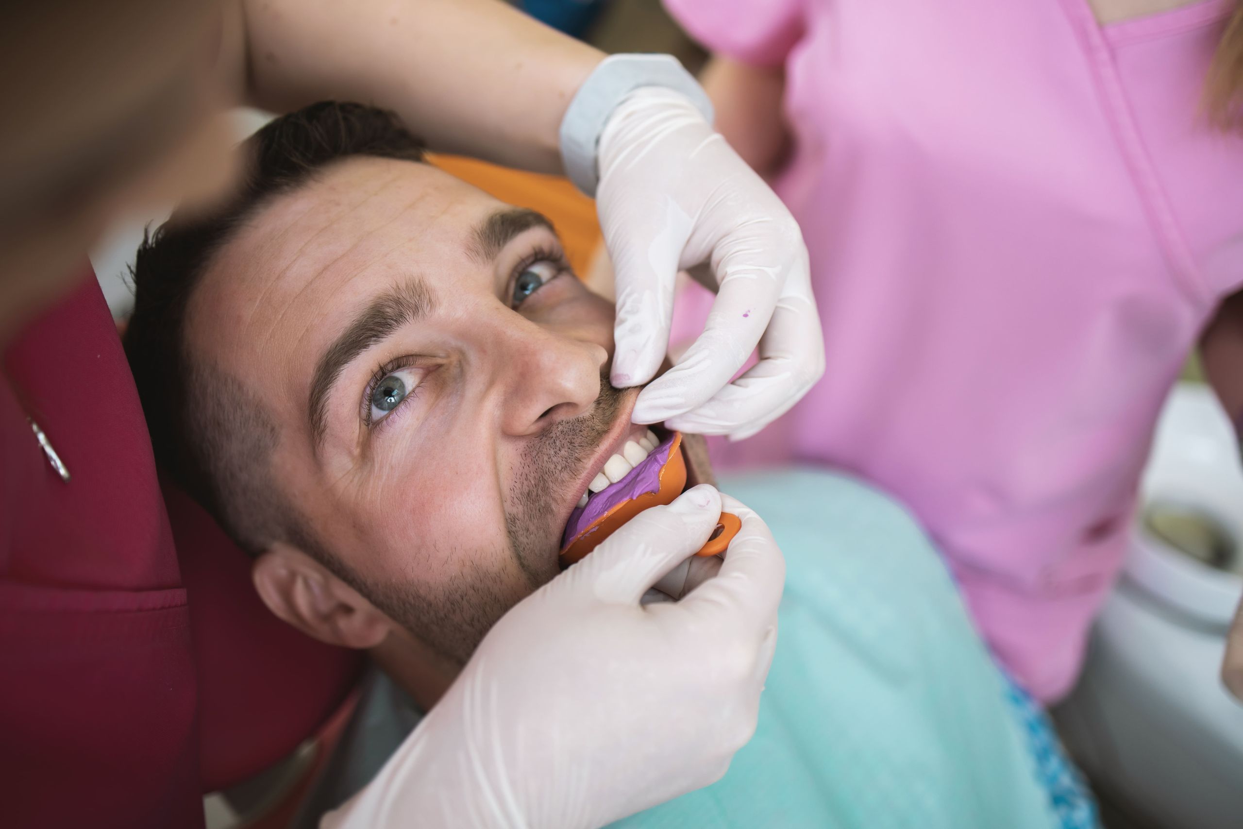
Traditional impressions use physical materials such as alginate or silicone to create a mold of the teeth. This process involves multiple steps, including material preparation, setting, removal, and often the creation of a physical model. Each of these steps can introduce variability through distortion, expansion, or handling, and may cause a gag reflex during the procedure.
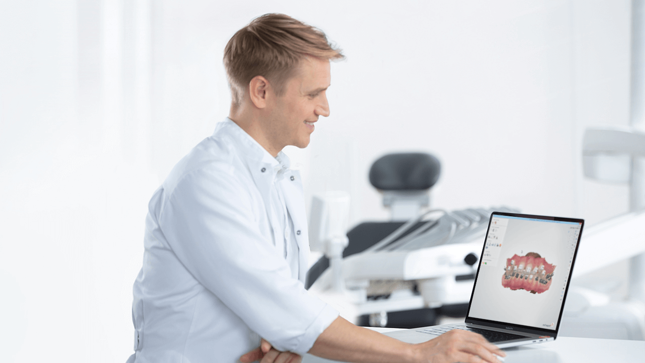
Digital impressions, in contrast, capture surface data directly using an intraoral scanner and convert it into a 3D digital model. This removes the need for physical materials and reduces the number of intermediate steps, allowing data to be processed, stored, and shared immediately.
These differences affect both clinical performance and workflow. Digital impressions generally improve patient comfort by avoiding impression trays and materials, reducing discomfort and minimizing the risk of gag reflex. They also reduce cross-infection risks associated with physical impression handling and support more efficient workflows by enabling faster data capture and transfer between clinics and laboratories, which can save time for both clinicians and patients 5.
Overall, both digital and traditional impression techniques remain clinically acceptable. However, digital workflows provide advantages in data handling, repeatability, and integration with CAD/CAM systems, making them increasingly used in modern dental practice.
What are digital impressions used for?
Digital impressions are used across multiple areas of dentistry to support diagnosis, treatment planning, and the design and manufacture of dental appliances and restorations. They serve as the starting point for both simple and complex procedures within digital workflows, including applications such as same-day restorative workflows.
Orthodontics
In orthodontics, digital impressions are used to create 3D models of the teeth for treatment planning and appliance design. These models help determine tooth position and guide the placement of braces, clear aligners, or other retainers.
They can also be used to monitor tooth movement over time and, in some cases, combined with additional imaging such as X-rays to support treatment decisions.
Dental implants
Digital impressions are used in implant workflows to support implant planning and positioning. This is often part of a prosthetically driven approach, where the final restoration guides the positioning of the implant.
In some cases, digital impression data is combined with other imaging data to create a complete treatment plan and to support the design of surgical guides.
Crowns, bridges, and restorations
Digital impressions are commonly used as the starting point for restorative treatments, including the design and fabrication of dental prostheses 2. They provide the data needed to design single-unit restorations such as crowns, as well as more complex cases such as bridges and full restorations.
The digital model is used in CAD/CAM systems to design and manufacture restorations with a precise fit.
Dentures and full-arch cases
Digital impressions are also used in digital denture workflows, including complete and partial dentures. They can be captured either directly from the patient or from a digitized physical model using an intraoral scanner.
Scanning fully edentulous patients can be more challenging due to soft tissue movement, but improvements in scanning technology and scan technique supported by research 6 have improved the feasibility of fully digital denture workflows.
Clinical and workflow implications of digital impressions
Digital impressions change how dental data is captured, handled, and used throughout the treatment process, supporting reliable treatment outcomes. By replacing physical impressions with digital data, they reduce the number of manual steps and enable more streamlined clinical workflows.
This has several practical implications:
- Reduced need for retakes, as scan data can be reviewed in real time and corrected immediately
- Faster communication between clinics and laboratories through digital file transfer
- Improved patient experience by eliminating impression materials and reducing chair time
- Digital storage of patient data, removing the need for physical model storage
- Integration with CAD/CAM systems for design and manufacturing of restorations and appliances
- More consistent and predictable workflows due to standardized data handling
- Improved efficiency across clinical and laboratory stages, as digital impressions can save time 5
These changes can improve coordination between clinical and laboratory stages, with some practices reporting measurable improvements in productivity based on improvements in workflow efficiency and productivity.
Common questions about digital impressions (FAQ)
What are digital impressions?
Digital impressions are 3D virtual models of a patient’s teeth and oral tissues created using an intraoral scanner. They capture surface data and convert it into a 3D digital file for diagnosis, treatment planning, and restoration design.
Are digital impressions more accurate than traditional impressions?
Digital impressions can achieve accuracy comparable to, and in some cases exceeding, traditional impressions, with studies reporting comparable or, in some cases, higher accuracy than traditional impressions 3. Accuracy depends on factors such as scanning technique, clinical conditions, and the system used.
Are digital impressions painful?
Digital impressions are generally not painful. They do not require impression materials or trays, which reduces discomfort and the risk of gag reflex.
Can digital impressions replace traditional molds?
Digital impressions can replace traditional molds in many clinical situations. However, certain cases, such as subgingival margins or fully edentulous scans, may still present challenges.
Are digital impressions suitable for all restorations?
Digital impressions are widely used for crowns, bridges, orthodontic appliances, implants, and dentures, including workflows such as digital denture fabrication. Suitability depends on the specific clinical case and scanning conditions.
What file formats are used for digital impressions?
Digital impressions are typically stored as 3D file formats such as STL or PLY. These formats are used in CAD/CAM systems for design and manufacturing.
How long does it take to take a digital impression?
The time required depends on the clinical case, the scanner, and operator experience. A study found that scanning time decreases significantly with operator experience 1. In many cases, a full-arch digital impression can be completed in under a minute, although this varies based on complexity and technique.
Summary: How digital impressions transform clinical workflows
Digital impressions are 3D virtual models of teeth and oral structures created using intraoral scanning technology. They replace physical impression materials by capturing surface data directly and converting it into a 3D digital format.
The process involves capturing optical data, processing it in real time, and generating a digital model for diagnosis, treatment planning, and restoration design. This reduces the number of manual steps and minimizes the risk of errors associated with physical materials.
By enabling faster data capture, immediate feedback, and seamless data transfer, digital impressions support more efficient communication between clinics and laboratories. They also improve patient experience and allow for more consistent and predictable workflows.
Evidence from multiple clinical studies indicates that digital impressions can achieve comparable or improved accuracy relative to conventional methods 7.
Overall, digital impressions play a central role in modern digital dentistry by supporting accurate, efficient, and integrated workflows.
Resources
- Róth I, Czigola A, Joós-Kovács GL, Dalos M, Hermann P, Borbély J. Learning curve of digital intraoral scanning - an in vivo study. BMC Oral Health. 2020 Oct 19;20(1):287. doi: 10.1186/s12903-020-01278-1. PMID: 33076894; PMCID: PMC7574448.
- Ting-Shu S, Jian S. Intraoral Digital Impression Technique: A Review. J Prosthodont. 2015 Jun;24(4):313-21. doi: 10.1111/jopr.12218. Epub 2014 Sep 14. PMID: 25220390.
- Nedelcu R, Olsson P, Nyström I, Rydén J, Thor A. Accuracy and precision of 3 intraoral scanners and accuracy of conventional impressions: A novel in vivo analysis method. J Dent. 2018 Feb;69:110-118. doi: 10.1016/j.jdent.2017.12.006. Epub 2017 Dec 12. PMID: 29246490.
- Fernandes A, Dua N, Herekar M. Corrective primary impression technique. Open Dent J. 2010 Apr 14;4:27-8. doi: 10.2174/1874210601004010027. PMID: 20502648; PMCID: PMC2874218.
- Yuzbasioglu E, Kurt H, Turunc R, Bilir H. Comparison of digital and conventional impression techniques: evaluation of patients' perception, treatment comfort, effectiveness and clinical outcomes. BMC Oral Health. 2014 Jan 30;14:10. doi: 10.1186/1472-6831-14-10. PMID: 24479892; PMCID: PMC3913616.
- Fang JH, An X, Jeong SM, Choi BH. Development of complete dentures based on digital intraoral impressions-Case report. J Prosthodont Res. 2018 Jan;62(1):116-120. doi: 10.1016/j.jpor.2017.05.005. Epub 2017 Jun 16. PMID: 28625663.
- 3Shape Scientific Research Overview Clinical Affairs, 2020: 34 clinical studies report statistical superior accuracy with TRIOS (2017-2019) Digital Versus Conventional Impressions in Dentistry: A Systematic Review, Chandran et al. 2019: Journal of Clinical & Diagnostic Research: 67% of (16 of 24) studies show digital impressions are more accurate (in microns) as conventional. 92% of (22 of 24) studies show the same level of clinical acceptability as conventional.