- Home
- Blog
- Digital dentistry
- How an intraoral scanner works
How intraoral scanners work: technology, process, and clinical implications
An intraoral scanner is a digital imaging device used to capture 3D data of teeth and oral structures. It works by projecting light onto the surfaces of the teeth and recording the reflected data. This data is processed into a digital 3D model used for diagnosis, treatment planning, and digital dental impression workflows in dentistry.
Understanding how intraoral scanners work is important for interpreting scan quality, accuracy, and clinical outcomes. The scanning process involves capturing optical data, processing it in real time, and converting it into a usable digital model. Each stage influences how accurately the final model represents the patient’s dentition.
A clear understanding of this process helps explain both the capabilities and limitations of intraoral scanning technology, including its role within digital dentistry workflows.
What is an intraoral scanner and what does it actually do?

In clinical use, an intraoral scanner captures a digital impression by scanning the surfaces of the teeth directly inside the patient’s mouth and is increasingly considered essential equipment in modern dental practice.
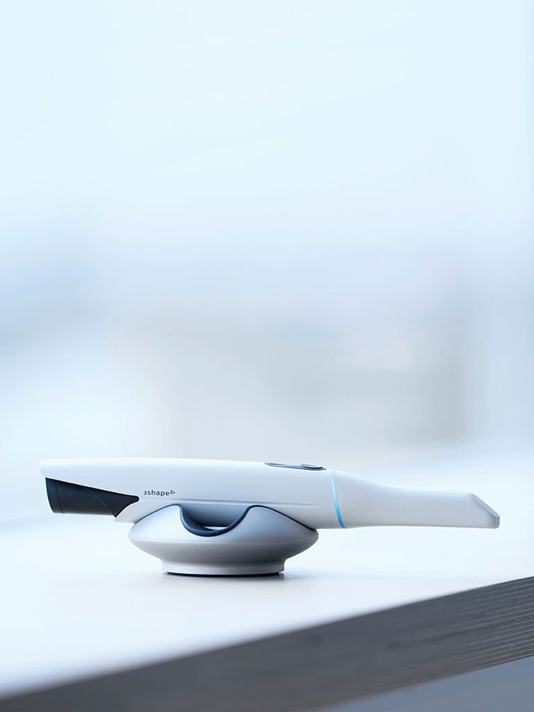
Modern systems are designed to be ergonomically suited for intraoral use, including devices with a compact intraoral scanner design.
The device is moved across the dentition, collecting visual data that is continuously processed into a 3D representation.
As the scan progresses, the system builds the model in real time, allowing the clinician to see the captured areas and identify any missing data.
The output of intraoral scanning is a digital dental impression that can be stored, analyzed, and shared between clinics and laboratories as part of a digital dentistry workflow.
How does an intraoral scanner work?
An intraoral scanner converts optical data from the surfaces of the teeth into a 3D digital model through a continuous scanning workflow. This process combines image capture, real-time data processing, and model generation into a single workflow.
During scanning, the device records optical data from multiple angles and positions. The software processes this data as it is captured, gradually building a complete digital representation of the dentition.
The final output is a digital dental impression that can be used for treatment planning, CAD/CAM design, and laboratory fabrication.
Process summary
An intraoral scanner projects light onto the teeth, captures the reflected data, processes it into a 3D surface model, and generates a digital dental impression for clinical use and laboratory workflows.
Step 1: Optical capture of tooth surfaces
The scanning process begins with optical capture. The intraoral scanner projects light onto the teeth and surrounding tissues, and imaging sensors record the reflected data.
Thousands of images are captured in rapid succession. These images represent different angles and positions of the tooth surfaces and form the basis for further processing.
Step 2: Real-time data processing
The captured images are processed by scanning software in real time. Ongoing software updates can improve processing performance, stability, and workflow efficiency. The system aligns and stitches the images together to create a continuous representation of the scanned area.
This process involves converting image data into point clouds and surface geometry, while continuously updating the model as additional scan data is captured. The software also filters out irrelevant data and ensures that only usable surface information is included.
Step 3: Creation of a digital dental impression
Once data has been captured and processed, the system generates a complete 3D digital model of the patient’s dentition.
This digital dental impression represents the geometry of the teeth and gingiva and can be used for diagnosis, treatment planning, and the design and manufacture of dental restorations within a digital workflow.
What technologies are used in intraoral scanners?
Intraoral scanners use optical systems and software to capture and process 3D data from teeth and oral structures. These technologies influence how surface data is captured, processed, and converted into a usable digital model.
Structured light and laser-based systems
Many intraoral scanners use structured light or laser projection. A known light pattern is projected onto the teeth, and sensors record how the pattern deforms across the surface.
This deformation allows the system to calculate depth and surface geometry, forming the basis of a 3D representation. Structured light systems are commonly used due to their ability to capture detailed surface information at high speed and resolution.
Confocal imaging and triangulation
Confocal imaging and triangulation are techniques used to determine depth from optical data.
Confocal imaging captures multiple images at different focal depths to isolate sharp surface data, while triangulation calculates the position of a surface point by measuring angles between the light source, object, and sensor.
Together, these methods enable scanners to build accurate depth maps from a sequence of 2D images, which are then combined into a 3D model.
Polarization and surface handling
Teeth present optical challenges due to their translucency and reflective properties. Light can scatter within the tooth or reflect unevenly from the surface, which can affect the accuracy of captured surface data.
Polarization techniques are used to filter reflected light and prioritize signals from the tooth surface. This improves the scanner’s ability to capture accurate surface data rather than subsurface noise, especially in enamel.
Powder vs non-powder systems
Earlier intraoral scanning systems required the application of powder to reduce reflections and improve surface contrast. The powder created a more uniform scanning surface but added an extra clinical step.
Modern intraoral scanners are typically powder-free and rely on advanced optics and software to manage surface properties. This simplifies the scanning process and improves patient comfort while maintaining consistent scan accuracy.
For a deeper explanation of surface behavior, see the optical characteristics of natural teeth.
How accurate are intraoral scanners?
Intraoral scanner accuracy refers to how precisely a digital scan represents the true shape and position of teeth and oral structures and this plays a critical role in treatment outcomes. Accuracy is typically assessed in terms of trueness (how close the scan is to the actual geometry) and precision (how consistent repeated scans are).
Clinical evidence indicates that intraoral scanners can achieve accuracy comparable to, and in some cases exceeding, traditional impression methods, depending on the indication and scanning conditions.
What affects intraoral scanner accuracy?
Several factors influence the accuracy of intraoral scanning, including both clinical conditions and how the scan is performed.
- Scanning technique and path used during image capture
- Accessibility of intraoral areas, especially interproximal and distal surfaces
- Presence of saliva, reflections, and soft tissue movement
- Optical properties of teeth, including translucency and surface texture
- Software algorithms used for image stitching and data processing
Managing these variables is important to ensure consistent and reliable scan results in clinical practice.
Digital scan accuracy vs traditional impressions
Digital and conventional impression methods differ in how accuracy is achieved and maintained throughout the workflow.
Traditional impressions rely on physical materials that can be affected by distortion, expansion, or handling during multiple manual steps. Each stage introduces potential variation in the final model.
Digital intraoral scanning captures data directly and reduces the number of steps required to produce a usable model. This minimizes the risk of material-related errors and allows for more consistent data handling.
Overall, both methods are clinically acceptable, but digital workflows can offer advantages in repeatability, data consistency, and integration with CAD/CAM systems.
Clinical implications: why the technology matters in practice
Intraoral scanning affects how dental treatments are planned and delivered across different clinical areas. By replacing physical impressions with digital data, it changes how information is captured, shared, and used throughout the treatment process.
Impact on restorative dentistry
In restorative dentistry, intraoral scanners provide accurate digital impressions that support the design and fabrication of restorations.
This affects restorative workflows in the following ways:
- Precise input for crowns, bridges, and implant restorations
- Accurate fit and potentially fewer adjustments during placement
- Better integration with CAD/CAM design and manufacturing systems
Impact on orthodontics
In orthodontics, intraoral scanning enables digital models that support diagnosis, treatment planning, and appliance production.
This affects orthodontic workflows in the following ways:
- Digital models for aligner design and case planning
- Easier monitoring of tooth movement over time
- Improved communication of treatment plans with patients
Workflow efficiency and chair time
Intraoral scanning can reduce the number of manual steps involved in impression-taking and data handling.
This affects clinical workflows in the following ways:
- May result in shorter chair time compared to traditional impression procedures
- Faster data transfer between clinic and laboratory
- Reduced need for impression materials and physical model handling
Clinical performance and workflow outcomes are often evaluated in independent research and clinical validation studies available in our library of clinical studies.
How different types of intraoral scanners compare
Intraoral scanners differ in how they capture, process, and interpret scan data, including how different systems are developed and refined over time. While all scanners are designed to create digital impressions, their performance can vary depending on the underlying technology and software.
The main differences include:
- Image capture and processing speed: Scanners vary in how quickly they capture images and convert them into usable 3D data
- Handling of difficult intraoral areas (such as interproximal surfaces and undercuts): Performance differs when scanning areas that are harder to access and capture accurately
- Software capabilities: The ability to process, stitch, and update scan data in real time varies across systems
- Consistency of scan results: Some systems produce more stable and repeatable results under different clinical conditions
These differences affect how reliably scanners perform in clinical situations and how easily complete digital impressions can be captured.
Comparing scanner models reveals how intraoral scanners differ in performance and usability.
Summary: How intraoral scanners convert light into digital dental models
Intraoral scanners capture optical data from teeth and oral structures by projecting light onto the surface and recording the reflected signal. This data is collected as a series of high-speed images during the scanning process.
The captured images are processed by software which identifies surface geometry, calculates depth, and combines the data into a structured 3D representation. Individual data points are organized into a mesh that forms a complete digital model of the scanned area.
The resulting digital impression can then be used for diagnosis, treatment planning, and integration with CAD/CAM systems for the design and production of dental restorations.
This process enables consistent data capture, reduces manual steps, and supports efficient digital workflows in clinical practice.
Explore the future of dental impressions with us